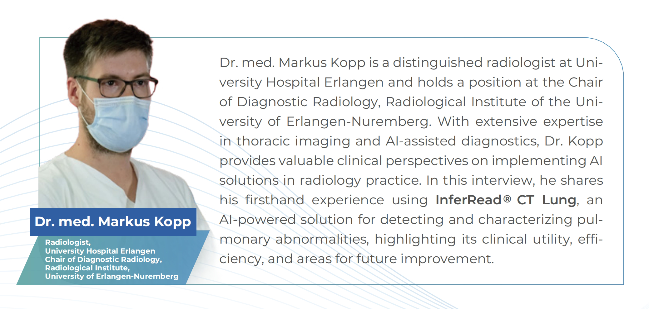
Introducing Dr. Med. Markus Kopp
User-Friendly Interface & High Diagnostic Accuracy
Dr. Kopp emphasizes that the AI platform is designed for seamless integration into a radiologist's workflow. "The interface is highly intuitive—most symbols are easy to understand, and within minutes, I was able to navigate the system efficiently." he notes. This ease of adoption ensures that clinicians can quickly incorporate AI into their daily practice without extensive training.
Beyond usability, the system demonstrates strong diagnostic performance. In one notable case, it "correctly identified small pulmonary nodules while also flagging a critical Pleural lesion characterized as potentially malignant." This capability is particularly valuable, as it expands diagnostic focus beyond the lungs. "Radiologists often concentrate on intrapulmonary findings, but AI helps us detect abnormalities in surrounding structures, like the chest wall, which might otherwise be overlooked." Dr. Kopp explains.
Efficient Processing Without Compromising Workflow
A key concern with AI adoption is whether processing delays could hinder productivity. However, Dr. Kopp finds the system’s five-minute turnaround time for CT scans aligns well with clinical demands. “For routine chest CTs, this is entirely reasonable—reports are typically generated within this timeframe anyway.” he says. The slight delay does not disrupt diagnostic workflows and ensures AI analysis is available when needed.
AI’s Strengths: Speed, Reconstruction, and Subtle Detection
Dr. Kopp highlights several advantages of AI in radiology:
· Rapid Anatomical Overviews: “Automatic reconstructions save significant time, giving us an immediate, clear view of anatomical regions.”
· Enhanced Detection of Subtle Abnormalities: The AI excels in identifying “small, easily missed subdural hematomas” in brain scans, suggesting similar potential for pulmonary diagnostics.

However, he acknowledges challenges, particularly false positives and the need for better integration into clinical decision-making. “How do we handle AI-flagged findings that don’t align with the radiologist’s report? This requires careful protocol development.”he notes.
The Future of AI in Radiology: Incidental Findings and Beyond
Looking ahead, Dr. Kopp sees significant potential in AI’s ability to flag incidental findings—abnormalities outside the primary area of focus. “If a spine CT incidentally captures part of the lungs, AI could identify nodules that a radiologist might miss.” he explains. Additionally, he anticipates advancements in radiomics for deeper lesion characterization.

Conclusion: A Valuable Tool with Room to Grow
Dr. Kopp’s experience underscores AI’s role as a supportive, rather than replacement, tool in radiology. Key takeaways include:
√ Intuitive design enables quick adoption.
√ High diagnostic accuracy for nodules and extrapulmonary lesions.
√ Minimal workflow disruption with fast processing.
√ Future potential in incidental findings and radiomics.
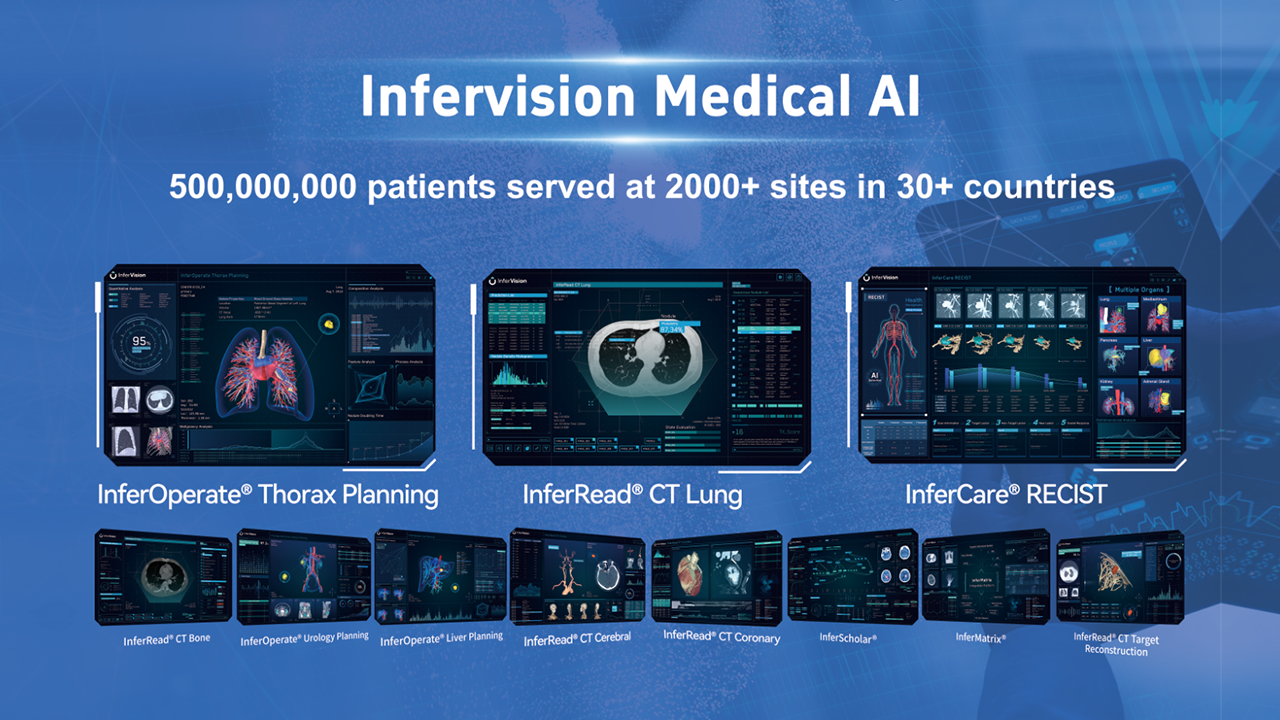
For clinicians considering AI integration, Dr. Kopp’s insights suggest that solutions like InferRead CT Lung ® can enhance diagnostic confidence—provided challenges like false positives and workflow integration are addressed. “AI won’t replace radiologists, but it will make us more thorough.” he concludes.