The Infervision research team has won 1st place in Pulmonary Artery Segmentation Challenge 2022 and Pulmonary Airway Tree Modelling Challenge 2022 held by MICCAI.
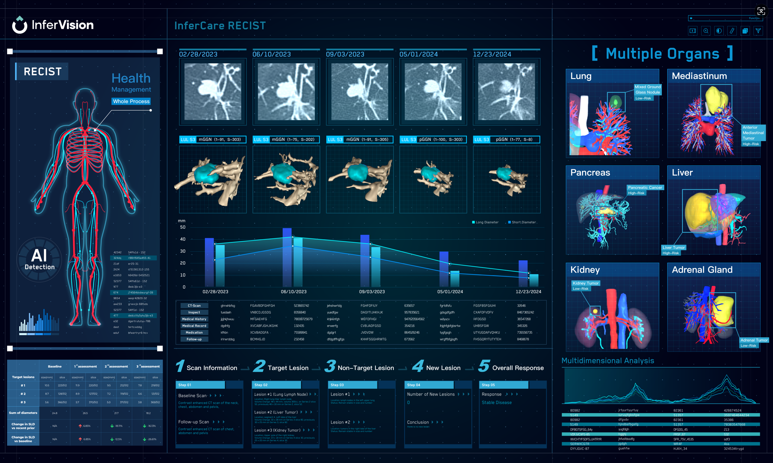
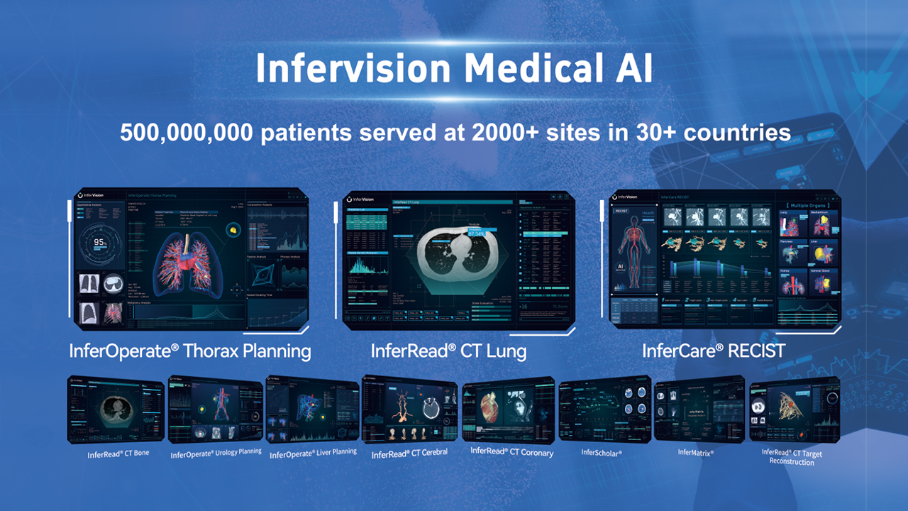
These two top-performing AI algorithms for segmentation empower the Inferoperate Thorax planning product, which focuses on chest 3D reconstruction. This new product can quickly and accurately segment anatomical structures such as pulmonary arteries and veins, tracheobronchial tubes, lung lobes and segments in lung CT, and assist doctors in diagnosis, treatment planning, and surgery planning.
👉About MICCAI
MICCAI is an academic conference organized by the Medical Image Computing and Computer-Assisted Intervention Society, which spans the fields of medical image computing (MIC) and computer-assisted intervention (CAI). It is recognized as the top international conference in the fields of medical imaging computing, medical robotics, artificial intelligence, assisted intervention, and computational biomedicine. In addition to the academic conferences, its annual challenges have attracted professionals, top universities, and renowned medical technology institutions worldwide.

👉About Pulmonary Artery Segmentation Challenge 2022
It is of significant clinical interest to study pulmonary artery structures in medical image analysis. One prerequisite step is to segment pulmonary artery structures from CT with high accuracy and low time-consuming. The segmentation of pulmonary artery structures benefits the quantification of its morphological changes for the diagnosis of pulmonary hypertension and thoracic surgery. However, due to the complexity of pulmonary artery topology, automated segmentation of pulmonary artery topology is a challenging task.
👉see the results here: https://lnkd.in/eeeh6uxr

👉About Pulmonary Airway segmentation Challenge 2022
Airway segmentation is a crucial step for the analysis of pulmonary diseases including asthma, bronchiectasis, and emphysema. The accurate segmentation based on X-Ray and computed tomography (CT) enables the quantitative measurements of airway dimensions and wall thickness, which can reveal the abnormality of patients with chronic obstructive pulmonary disease (COPD). Besides, the extraction of patient-specific airway models from CT images is required for navigation in bronchoscopic-assisted surgery. Due to the fine-grained pulmonary airway structure, manual annotation is however time-consuming, error-prone, and highly relies on the expertise of clinicians.
👉see the results here: https://lnkd.in/eAsg_P4c