OVERVIEW
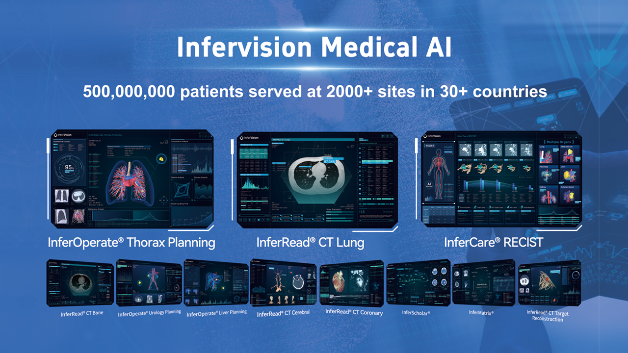
The Hepatobiliary Surgery Department at The Second Affiliated Hospital of Soochow University successfully employed InferOperate's advanced 3D reconstruction model in the management of a complex recurrent liver cancer case in a 60-year-old female patient.
Patient Profile:
- Age/Gender: 60-year-old female
- Initial Diagnosis: Liver cirrhosis and ascites (diagnosed four years ago)
- Previous Treatments: Laparoscopic liver cancer resection and Transarterial Chemoembolization (TACE)
CASE PROGRESSION
- June 2021: MRI scans revealed abnormal liver lesions. The patient was diagnosed with hepatocellular carcinoma (HCC) and underwent laparoscopic resect ion.
- October 2023: Follow-up MRI indicated a possible recur rence of HCC. A partial hepatectomy was per formed successfully.
- Post-Surgery: Enhanced CT scans detected new lesions, suggesting further recurrence.
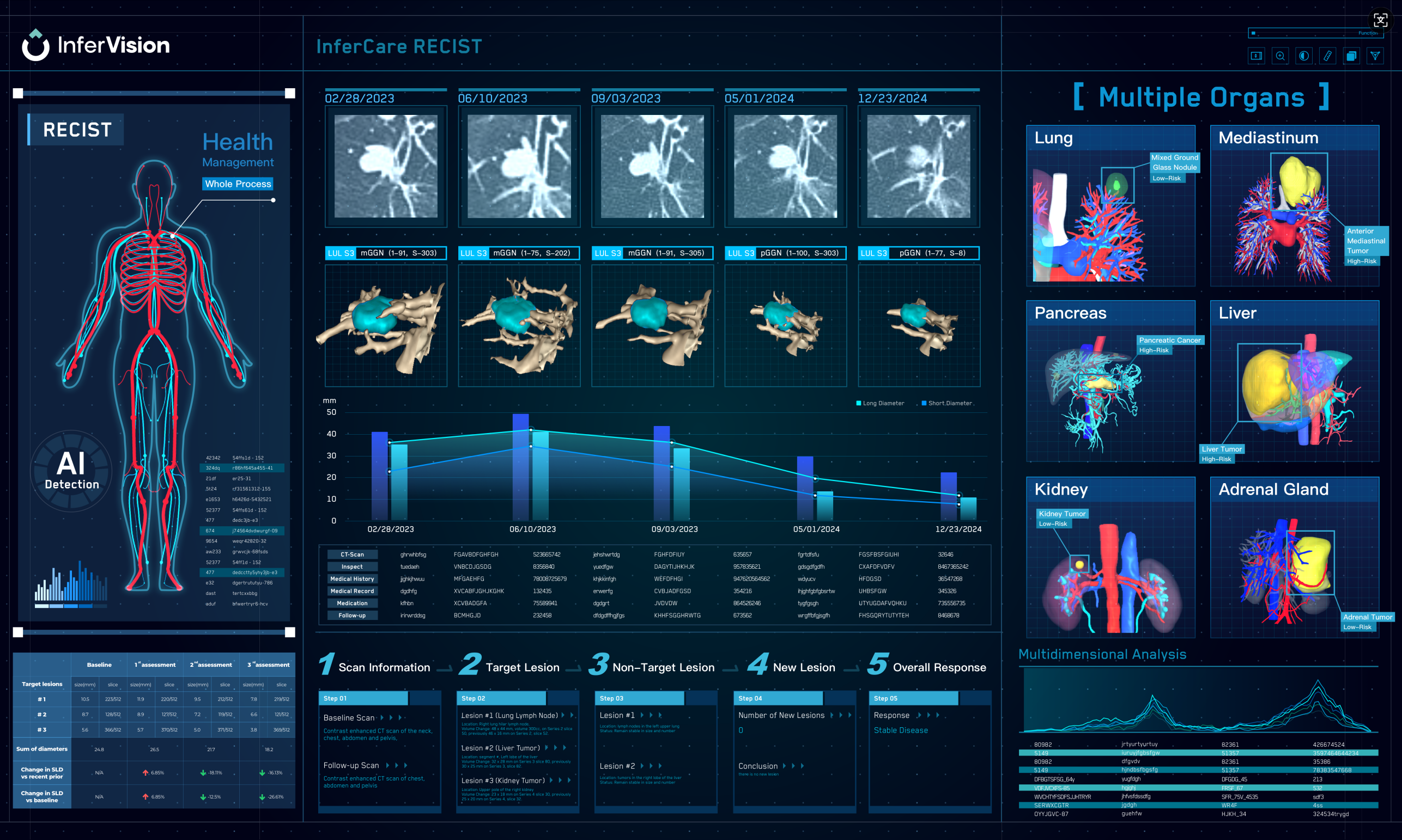
3D RECONSTRUCTION APPLICATION
Volume Calculation:
Traditional imaging was inadequate for assessing the remaining liver volume. InferOperate’s 3D reconstruction revealed the left liver volume to be 38.1% and the remaining liver volume at 61.9%, enabling the surgical team to proceed with a confident left hepatectomy.
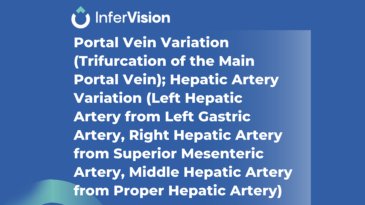
Tumour Localization:
The 3D model accurately mapped liver segments and their proximity to critical blood vessels, guiding the surgical plan for precise tumour excision.
Intraoperative Guidance:
The integration of 3D images with intraoperative ultrasound allowed for real-time assessment of tumour size, depth, and its relationship with surrounding vasculature, thereby enhancing surgical accuracy.
SURGICAL PROCEDURE
- A 25 cm "reverse L-shaped" incision was made to expose the left liver lobe.
- Intraoperative ultrasound and 3D reconstruction confirmed the tumour's precise location.
- A left hepatectomy was performed with meticulous dissection and vessel management.
- The patient recovered well post-surgery, with minimal bleeding and no transfusion required.
CONCLUSION
The case exemplifies the critical role of 3D reconstruction in complex liver surgeries, particularly in accurate tumour localization and volume assessment, ultimately leading to better patient outcomes.
✓ Reduced clinican workload and reconstruction time
✓ Improved surgical outcomes
Ready to transform your surgical planning?
👆Contact us today to schedule a demo with one of our team!
👆Click here to download the full case of the week!